|
Concept of AXON . The object of the simulation is a reaction of a
nerve (axon) in a muscle. The simulation has 3 graphs: - black (1) graph shows the impulse line; - green (2) and red (3) graphs show the reaction
of the muscle. There are 2 types of nerve (axon) reaction -
normal reaction and suppressed reaction. When the first impulse starts
there is a normal nerve reaction. After a short period there comes the
second impulse . But nerve (axon) does not react in normal way. Reaction
to the second impulse is very low. This phenomena can be explained in term of
weariness of nerve. An axon itself is a kind of electric cable that
connects the impulse generator (nerve-cell body) and the object (muscle).
A nerve has very complex construction and process.
The human body is made up of trillions of cells. Cells of
the nervous system, called nerve cells or neurons,
are specialized to carry "messages" through an electrochemical
process. The human brain has about 100
billion neurons. Neurons come in many different shapes and sizes. Some of the smallest
neurons have cell bodies that are only 4 microns wide. Some of the
biggest neurons have cell bodies that are 100 microns wide. Neurons
are similar to other cells of the body because:
However, neurons differ from other
cells in the body because:
Pic.1. There are several differences between axons and dendrites:
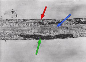
The
pic.2. shows a longitudinal section of a nonmyelinated nerve axon. There
is a longitudinal section of a mitochondrion
visible. The most striking feature of this electron micrograph is the microtubules.
They stretch all the way down the nerve axon and are instrumental in
axonal growth and transport. Also evident are the plasma
membrane of the axon, sometimes called the axolemma, and the
axoplasm, the cytoplasm of the axon.
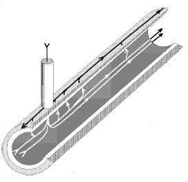
Pic.2. There
is a portion of an axon immersed in a conductive solution in the pic.3.
There is an electrode penetrating the surface which is supplying current
with respect to another electrode located outside, and the current
circulation is pictured by a few lines of flow. If the membrane were a
perfect insulator, no current would flow towards the external electrode.
But due to the finite permeability of the axon, some current will
circulate longitudinally in the axon interior, and exit through the
surface membrane to be finally collected by the external electrode. It
is clear then that due to the current circulation and the resistivity of
the axoplasm, there will be voltage drops along
the axon and none of the two sides of the membrane will be isopotential.
Pic.3. Neurons send messages through an electrochemical
process. This means that chemicals result in an electrical signal.
Chemicals in the body are "electrically-charged" -- when they
have an electrical charge, they are called "ions."
The important ions in the nervous system are sodium and potassium (both
have 1 positive charge, +), calcium (has 2 positive charges, ++) and
chloride (has a negative charge, -). There are also some negatively
charged protein molecules. It is also important to remember that nerve
cells are surrounded by a membrane that allows some ions to pass through
and blocks the passage of other ions. This type of membrane is called semi-permeable.
When a neuron is not sending a signal, it is said to be "at rest." When a neuron is at rest, the inside of the neuron (pic.4) is negative relative to the outside. Although the concentrations of the different ions attempt to balance out on both sides of the membrane, they cannot because the cell membrane allows only some ions to pass through channels (ion channels). At rest, potassium ions (K+) can cross through the membrane easily. Also at rest, chloride ions (Cl-)and sodium ions (Na+) have a more difficult time crossing. The negatively charged protein molecules (A-) inside the neuron cannot cross the membrane. In addition to these selective ion channels, there is a pump that uses energy to move 3 sodium ions out of the neuron for every 2 potassium ions it puts in. Finally, when all these forces balance out, and the difference in the voltage between the inside and outside of the neuron is measured, you have the resting potential. The resting membrane potential of a neuron is about -70 mV (mV=millivolt) - this means that the inside of the neuron is 70 mV less than the outside. At rest, there are relatively more sodium ions outside the neuron and more potassium ions inside that neuron.
Pic.4. So the resting potential indicates what is happening with the neuron at rest. The action potential indicates what happens when the neuron transmits information down an axon, away from the cell body (pic.5). Neuroscientists use other words, such as a "spike" or an "impulse" to describe the action potential. The action potential is an explosion of electrical activity that is created by a depolarizing current. This means that some event (a stimulus) causes the resting potential to move toward 0 mV. When the depolarization reaches about -55 mV a neuron will fire an action potential. This is the threshold. If the neuron does not reach this critical threshold level, then no action potential will fire. Also, when the threshold level is reached, an action potential of a fixed sized will always fire...for any given neuron, the size of the action potential is always the same. There are no big or small action potentials in one nerve cell - all action potentials are the same size. Therefore, the neuron either does not reach the threshold or a complete action potential is fired - this is the "ALL OR NONE" principle.
The
"cause" of the action potential
is an exchange of ions across the neuron membrane. A stimulus first
results in the opening of sodium channels. Because there are a lot more
sodium ions on the outside, and the inside of the neuron is negative
relative to the outside, sodium ions rush into the neuron. Remember,
sodium has a positive charge, so the neuron becomes more positive and
becomes depolarized. It takes longer for potassium channels to open.
When they do open, potassium rushes out of the cell, reversing the
depolarization. Also at about this time, sodium channels start to close.
This causes the action potential to go back toward -70 mV (a
repolarization). The action potential actually goes past -70 mV (a
hyperpolarization) because the potassium channels stay open a bit too
long. Gradually, the ion concentrations go back to resting levels and
the cell returns to -70 mV. |